baby chest x ray technique
When the chest radiograph also includes the abdomen look out for the umbilical clip. During the examination an X-ray machine sends a beam of radiation through the chest and an image is recorded on special film or a computer.

Chest X Ray Of A 6 Month Old Child With An Icd The Active Can Is Download Scientific Diagram
It is often the first type of imaging used to identify sources of pain evaluate traumatic injuries and locate a foreign body.

. The process generates a white and black image of your kids chest by projecting a small radiation beam through the chest. Chest x-ray is the most commonly used imaging exam for evaluating the chest. These are plastic clips used to clamp the umbilicus before it is cut at birth.
Chest AP erect in chair 180 cm. 851a a Use automatic exposure control 500 speed for chestabdomen else 400 speed at specified kVp when practical. X-rays are a diagnostic test that uses invisible electromagnetic energy beams to produce images of internal tissues bones and organs onto film.
A chest X-ray is a safe and painless test that uses a small amount of radiation to take a picture of a persons chest. This is partly due to modesty but also due to fear. The developed AI algorithm not only predicts whether the CXR has.
A chest X-ray also helps diagnose meconium aspiration. Lateral cervical spines are taken at 150 cm. A standard chest x-ray includes an anteroposterior AP supine projection of the chest.
Aerationofthenormalneonatallungisvirtuallycomplete within two or three respiratory cycles after birth and the lung fields should appear symmetrically aerated on the initial X-ray with the diaphragms lying at the level of. This image includes organs and structures such as the heart lungs large blood. Supporters see fewerno ads.
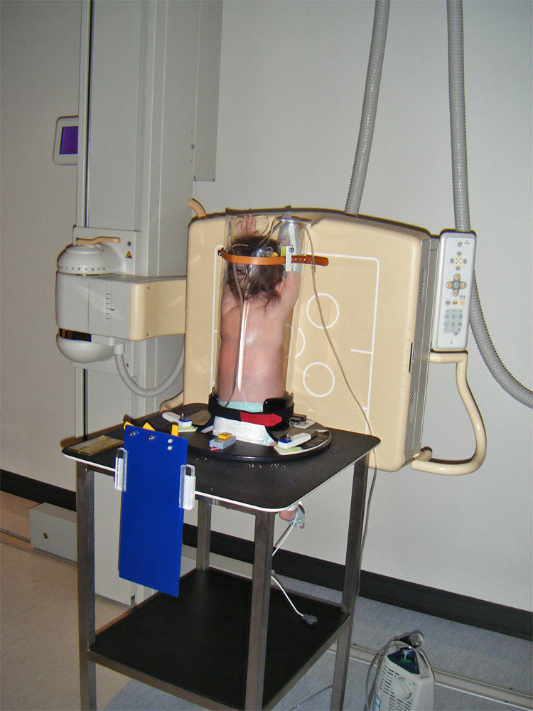
The researchers from IIT Jodhpur have developed an automated Artificial Intelligence AI solution for COVID-19 prediction from chest X-raysThe experiment was performed with more than 2500 chest X-ray images and achieved about 9680 per cent sensitivity. Up to 10 cash back The radiographer inside the cubicle positions the bed close to the glass door and places the digital detector behind the patient for an erect anteriorposterior chest X-ray before stepping away from the patient during the exposure a while the radiographer outside the cubicle positions the X-ray tube head close to the glass door and steps laterally. Erect chest X-rays are taken at 180 cm.
Full legfull spine imaging is performed at 180 cm using CR. The aim of this study was to develop and validate a prediction model to estimate the probability for a normal chest x-ray in children with RSV infection. This is sufficient for diagnosis of respiratory problems however it is necessary to include 23 of the abdomen in order to clarify that other factors such as abdominal gas are not causing the respiratory symptoms and also to check the position of umbilical venous and arterial.
Head is straight and chin raised out of the field of view. Most neonatal chest X-rays are AP films unless the baby is made to lie prone Lucency of soft tissue shadow - darker the soft tissue more is the exposure Ease of visibility of retrocardiac vertebrae if the retrocardiac vertebrae are easily seen the film is over exposed Relative lucency of lung fields. Pediatric Chest Screen 70-80 DIGITAL OPTIMUM kVp Universal CR Technique Chart using a standard 21 LgM Part View kV mAs kV mAs kV mAs Abdomen AP Grid 85 10 -15 85 20 - 25 85 30 - 40 Ankle AP 70 18 70 2 70 25 Ankle Obl 70 16 70 18 70 22 Ankle Lat 70 15 70 16 70 2 Chest -Adult AP 400 - tt -72 85 2 - 25 85 32 - 4 90 5 - 64.
IIT Researchers Develop Technique To Diagnose COVID-19 Using Chest X-Rays The experiment was performed with more than 2500 chest X-ray images and achieved about 9680 per cent sensitivity. The presence of meconium in the amniotic fluid is key to the diagnosis. The normal neonatal chest X-ray.
Shoulders touching the detector to ensure no rotation 1. A chest x-ray is one of the most common and safe diagnostic tests. A chest radiograph for a 12-year-old female is an embarrassing ordeal.
X-rays are used throughout the body. Patient Position Infant. All distal extremity exposures are taken at 110115 cm SID.
X-ray exams are used to help diagnose a wide variety of injuries and illnesses in children. Ideally shoulders rolled anteriorly to remove scapulae off chest wall. Immobilize legs with Ace bandage or tape and sandbags.
However all children are modest to some degree about having their genitals or backsides exposed after ages 4 to 5. Exposure chart was developed with two distinct groups of exposure. Center Image receptor to Central Ray.
A chest x-ray is frequently performed in infants with LRTI caused by RSV. The process generates a white and black image of your kids chest by projecting a small radiation beam through the chest. Immobilize infants arm above the head or use stockinette ace bandage tape or sandbags.
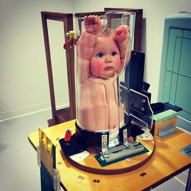
In contrast most 12-year-old males have little modesty about their chests. 901a a Use automatic exposure control 500 speed for chestabdomen else 400 speed at specified kVp when practical. The chest radiograph is one of the most commonly requested radiographic examinations in the assessment of the pediatric patientDepending on the patients age the difficulty of the examination will vary often requiring a specialist trained radiographer familiar with a variety of distraction and immobilization techniques.
However if the child struggles with this position they can hug the detectorbucky. Use gonadal shielding if possible. The umbilical stump remains in situ for approximately 1-2 weeks and.
Optimisation strategies body exposures for head trunk humerus femur and. Chest PAAP erect 180 cm.
Ce4rt Guide For X Ray Techs To Immobilize Pediatrict Patients

Pediatric Chest Ap Erect View Radiology Reference Article Radiopaedia Org

Neonate Chest Supine View Radiology Reference Article Radiopaedia Org

Pediatric Chest Horizontal Beam Lateral View Radiology Reference Article Radiopaedia Org
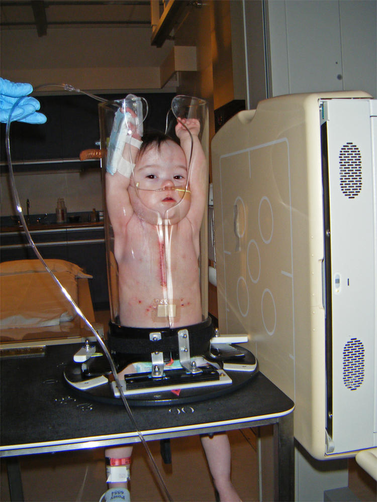
Paediatric Chest Immobilisation Devices Wikiradiography
Paediatric Chest Immobilisation Devices Wikiradiography

Pediatric Chest Supine View Radiology Reference Article Radiopaedia Org
Pedia Poser For Xray Imaging
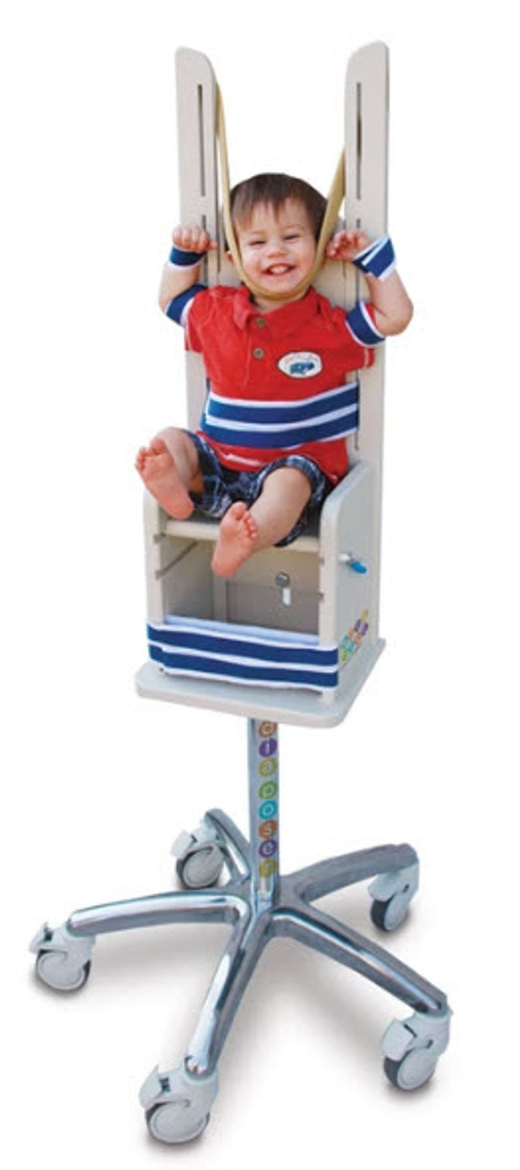
Pedia Poser For Xray Imaging